Arterial Sonogram Legs

Leg Arterial Normal Ultrasoundpaedia

Leg Arterial Normal Ultrasoundpaedia

Leg Artery Doppler Ultrasound And Ankle Brachial Index Abi Cremorne Radiology

A Arterial Ultrasound Of Right Popliteal Artery Demonstrating Complete Download Scientific Diagram
Doppler Ultrasound Exam Of Arm Or Leg Purpose Results And More

Leg Arterial Normal Ultrasoundpaedia
Each leg contains a number of large arteries the biggest of which is the femoral artery.
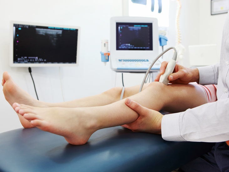
Arterial sonogram legs. Your doctor may recommend an arterial duplex ultrasound if they suspect an artery is narrowed or blocked reducing blood flow to your arms or legs. This exam checks to see if you have any blockages in the arteries that take blood to the legs by taking blood pressures from various locations. 5 the popliteal artery.
Arterial evaluation localizes sites of obstruction by segmental pressures obtained by Doppler signal assessment using pressure cuffs. An ultrasound of your leg may be requested to check for arterial blockage a blood clot inside a blood vessel or an injury to a blood vessel. A Doppler ultrasound study a technique that evaluates blood flow through a blood vessel is usually part of this exam.
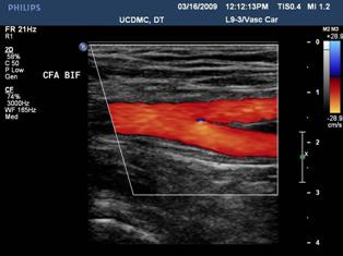
Narrowing of your vessels that may be causing leg pain when walking. A Doppler ultrasound uses sound waves to produce images that highlight blood flow in. Third arteries have visible walls and sometimes have calcified plaques on.
An arterial exam of the legs is a non-invasive test using blood pressure cuffs. When performing duplex ultrasound the technologist will begin at the common femoral artery CFA and move distally down the leg to thoroughly evaluate all the main arteries. Duplex ultrasound of the leg arteries shows blockages that can cause leg pain with walking.
This test is done as the first step to look at arteries and veins. Second arteries are smaller than veins. No radiation dyes or needles are used for this exam.
Carotid duplex ultrasound looks at the carotid artery in the neck. What Happens During Arterial Duplex Ultrasound Lower Legs. This clinical guide will show you how to identify symptoms perform a basic arterial duplex and ankle-brachial index ABI and interpret the findings so that you can give immediate results to your patients.

Doppler Ultrasound Of An Artery Northshore

Doppler Waveform In Femoral Artery Before And After The Exercise On Ultrasound Google Search Ultrasound Sonography Vascular Ultrasound Medical Ultrasound

Tips For Locating Lower Extremity Arteries On Ultrasound Medmastery
Peripheral Arterial Duplex Scanning Vascular Center Uc Davis Health
Ultrasonography

Vascular Ultrasound Lecture How To Detect An Occlusion Of The Superficial Femoral Artery Youtube

Basic Anatomy Of The Lower Extremity Arteries Medmastery
Ultrasound Assessment Of Lower Extremity Arteries Radiology Key

Leg Arterial Normal Ultrasoundpaedia

